Protist Lab:
objective- to observe prepared slides and learn about different protist's structures
results-
hypothesis- If we can figure out how to focus the microscope properly, then we can observe and learn about protists.
materials-
light microscope
prepared slides
micropipet
cover slips/slides
protozoan culture
procedure-
1. observe prepared slides with 4x, 10x, 100x objectives
2. make wet mount slide using drop of protozoan culture and observe at each objective
3. clean/dry cover slips and return them
conclusion:
My hypothesis was correct in that we figured out how to focus the microscope. Two things that we can improve in the future are focusing more throughout the lab and trying to focus each time completely correct, but we did improve our microscope techniques.
objective- to observe prepared slides and learn about different protist's structures
results-
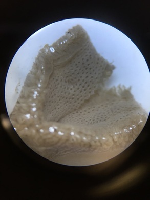
- structure for Euglena- fuzzy dots that look like small paramecium
- structure for Amoeba- shaped like stars, blobs, not particular, colored pink, yellow, green, blue
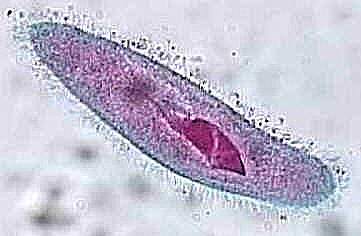
- structure for paramecium- little pink things, kind of look like fish, or an eye
- vortecella- looks like a red branch, and seems like a crack
- protist mix- looks like a mixture of amoeba and paramecium
- live paramecium- looks longer and skinner, but same idea as dead ones
- live protozoa- we couldn't find/see anything
hypothesis- If we can figure out how to focus the microscope properly, then we can observe and learn about protists.
materials-
light microscope
prepared slides
micropipet
cover slips/slides
protozoan culture
procedure-
1. observe prepared slides with 4x, 10x, 100x objectives
2. make wet mount slide using drop of protozoan culture and observe at each objective
3. clean/dry cover slips and return them
conclusion:
My hypothesis was correct in that we figured out how to focus the microscope. Two things that we can improve in the future are focusing more throughout the lab and trying to focus each time completely correct, but we did improve our microscope techniques.